
Stilla Technologies Digital PCR System
naica®Crystal Digital PCR System
NaicaTMCrystal Digital PCR System
Azure Biosystems Real-Time PCR Systems

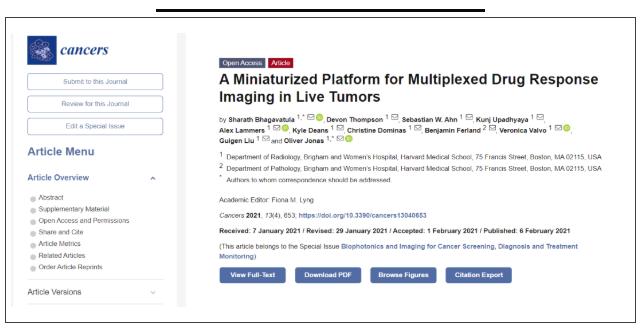
In 2020, the number of new cancers reached 4.57 million, and the number of deaths due to cancer was even more than 3 million. Therefore, more than effective programs and more reasonable cancer treatments were discovered. Currently available measures of drug response, such as tumor shrinkage, often take weeks to months to manifest and are not always representative of a tumor’s dynamic sensitivities. Implantable microdevices (IMD) have been recently developed to deliver microdoses of chemotherapeutic agents locally into confined regions of live tumors; the tissue can be subsequently removed and analyzed to evaluate drug response. This method has the potential to rapidly screen multiple drugs, but requires surgical tissue removal and only evaluates drug response at a single timepoint when the tissue is excised. The article described a “lab-in-a-tumor” implantable microdevice (LIT-IMD) platform to image cell-death drug response within a live tumor, without requiring surgical resection or tissue processing.

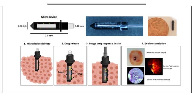
LIT-IMD combines miniature imaging probes, implantable miniature devices, and live cell detection technology to insert them into live tumors. IMD delivers multiple drug microdoses to discrete locations, and at the same time, it releases locally A small amount of fluorescent cell death detection agent (propidium iodide PI, PI binds to double-stranded DNA, but can only enter cells that have destroyed the cell membrane; therefore, it accumulates in late apoptotic and necrotic cells that lose membrane integrity), diffuse To the tissue exposed to the drug and accumulate at the site of cell death. The integrated miniaturized fluorescent imaging probe images each area to assess drug-induced cell death.

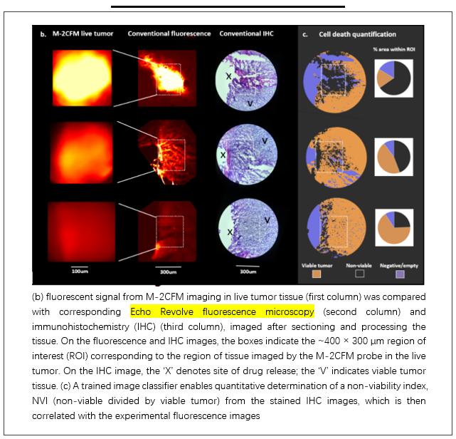
LIT-IMD was used to evaluate multiple drug responses in a mouse tumor model at 8 hours,Current gold-standard methods to evaluate cell death include conventional benchtop fluorescence imaging of propidium iodide and immunohistochemistry (IHC) analysis.,The researchers compared tumor imaging with the gold standard fluorescence microscope and histopathology, and used the Echo Revolve fluorescence microscope for standard imaging. The LIT-IMD imaging results are obviously correlated with the Echo Revolve fluorescence microscope images.

(b) fluorescent signal from M-2CFM imaging in live tumor tissue (first column) was compared with corresponding Echo Revolve fluorescence microscopy (second column) and immunohistochemistry (IHC) (third column), imaged after sectioning and processing the tissue. On the fluorescence and IHC images, the boxes indicate the ~400 × 300 µm region of interest (ROI) corresponding to the region of tissue imaged by the M-2CFM probe in the live tumor. On the IHC image, the ‘X’ denotes site of drug release; the ‘V’ indicates viable tumor tissue. (c) A trained image classifier enables quantitative determination of a non-viability index, NVI (non-viable divided by viable tumor) from the stained IHC images, which is then correlated with the experimental fluorescence images
This platform is the first fully integrated platform that can be used to evaluate multiple chemotherapy responses in situ. During the development of the imaging platform, the Echo Revolve fluorescence microscope helped developers determine the platform-related shooting effects and instrument performance through good imaging effects and parameters, and helped optimize and perfect the platform performance.
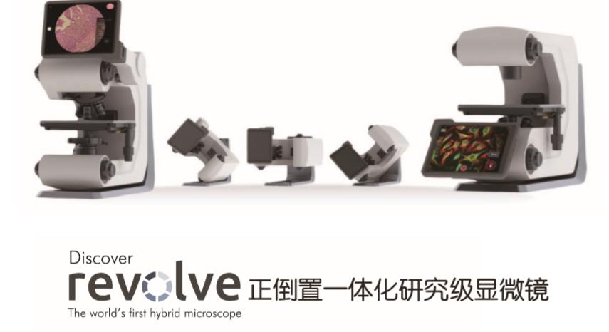
Echo Revolve microscope

☑ Retina display:Comparing eyepieces to human eyes。
☑ Full field observation: Clearer and more convenient。
☑ Multichannel fluorescence:Up to 4 EPI fluorescent channels,Multicolor fluorescence microscopic analysis can be done quickly and easily without darkroom.
☑ Automatic operation:The switch between the camera and the fluorescence channel is controlled by iPad Pro, which realizes fully automated operation。
☑ IOS-based Echo app:IOS-based Echo App is a professional microscope software developed in cooperation with the Apple team。
☑ Exquisite craftsmanship, high-end quality:Achieve extraordinary performance。
