Stilla Technologies Digital PCR System
naica®Crystal Digital PCR System
NaicaTMCrystal Digital PCR System
Azure Biosystems Real-Time PCR Systems


Introduction
Western blotting was first described by three groups in 1979. Since then numerous refinements to the basic technique, reagents used and imaging technologies have massively broadened the usage of Western blotting, making it a cornerstone protocol in life sciences today.
However, the general workflow of a Western blot has remained the same throughout this time. Samples are isolated and protein is extracted and standardized.Proteins are then separated by gel electrophoresis,transferred and probed with primary and secondary antibodies before being visualized. With numerous steps, Western blotting as a protocol requires a large investment in equipment, training, reagents and,perhaps most importantly, time.
A standard Western blot protocol takes two days to work from sample to image. Therefore, when analyzing a large number of samples or performing optimizations,it is easy to see how Western Blotting can quickly become a bottleneck in your research workflow.
Enter in-cell Western blotting – a radical rethinking of the standard protocol coupling the ability to accurately quantify intracellular proteins from Western blotting with the repeatability, quick turnaround and highthroughput of an ELISA. Cells are grown on sterile plates,before being fixed and permeabilized in situ. Subsequent labelling is performed as normal with a specific primary antibody followed by an isotype specific fluorescent conjugated secondary antibody. Conjugates in the nearinfrared (NIR) spectrum are commonly used to reduce auto-fluorescence and noise typically associated with tissue culture plastics.
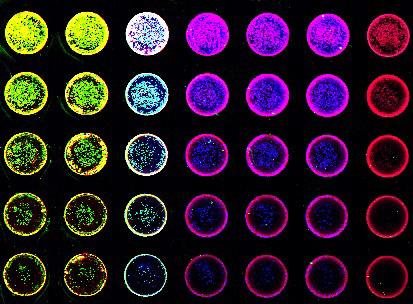
Figure 1. Representative image of an in-cell Western blot.
The Azure Biosystems Sapphire™ Biomolecular Scanner is specifically designed to allow for visualization in two NIR channels (658 nm and 785 nm) as well as visible fluorescent wavelengths (488 nm and 520 nm), allowing for multiplex imaging within wells. This multiplex capacity allows for the assessment of multiple proteins of interest in a single well which is useful for looking
at total and phosphorylated versions of a protein or to detect an in-well standard to allow for quantification across a plate.
With a laser based excitation system using photomultiplier tube (PMT) and avalanche photodiode (APD) detectors, signal selectivity, sensitivity and speed of imaging achievable by the Sapphire make it an ideal choice for in-cell Western blotting.
Materials and Methods
Culture cells
HeLa cells were serially diluted and seeded into a sterile 96 well tissue culture plate at a volume of 0.2 mL per well, and grown until approximately 80% confluent.All wells were fixed and permeabilized using 100% methanol for 15 minutes at room temperature.
In-cell Western blotting
Following fixation and permeabilization, cells were rinsed in PBS, blocked with 1% fish gelatin in PBS for one hour at room temperature, then probed for alpha-tubulin and beta-actin overnight at 4°C. Samples were washed three times with PBS prior to incubation with AzureSpectra 550 and AzureSpectra 800 conjugated secondary antibodies for 60 minutes.
Secondary antibodies were incubated along with RedDot™1 Nuclear Stain to normalize for total cell number. Plates were washed as previously described before imaging.
Imaging
After washing the plate was imaged on the Azure Biosystems Sapphire™ Biomolecular Scanner using the Plate focus setting.
Results and Conclusions
In this note we demonstrate the ability to quantify intracellular proteins in situ using the Azure Biosystems Sapphire™ Biomolecular Scanner. HeLa cells were grown in a 96-well plate probed with alpha-tubulin and beta-actin antibodies followed by isotype appropriate secondary antibodies conjugated with AzureSpectra 550 and AzureSpectra 800 respectively. All wells were
incubated with RedDot™1 Nuclear Stain to normalize for total cell number. Images were acquired using an Azure Biosystems Sapphire™ Biomolecular Scanner and are displayed in Figure 2. The representative image shown demonstrates the high level of sensitivity and specificity achievable.
Performing in cell Western blotting on 96 well plates allows for accurate measurement of intracellular proteins expression in situ; and provides a high throughput methodology to assess multiple stimulations,end-points, proteins of interest and replicates on a
single plate. By using NIR antibodies and the Azure Biosystems Sapphire™ Biomolecular Scanner the potential for in well multiplex analysis also exists offering further improvements to throughput.
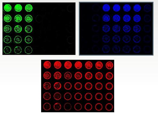

Figure 2. A serial dilution of HeLa cells were seeded into a 96-well plate, cultured, fixed and permeabilized. A) Columns 1-3 were probed for beta-Actin using AzureSpectra 550 (green). B) Columns 3-6 were probed for Tubulin using AzureSpectra 800 (blue). C) The entire plate was stained with RedDot1 Nuclear Stain as a normalization control (red). D) The individual channels were scanned simultaneously then combined into a single composite image using the Sapphire Capture Software.